[1] Fetsch PA, Marincola FM et al. (1999): Melanomaassociated antigen recognized by T cells (MART-1): the advent of a preferred immunocytochemical antibody for the diagnosis of metastatic malignant melanoma with fine-needle aspiration. Cancer 87(1): 37-42.
[2] Kageshita T, Kawakami Y et al. (1997): Differential expression of MART-1 in primary and metastatic melanoma lesions. J Immunother. 20(6): 460-5.
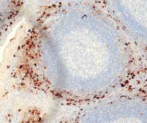
[3] Orosz Z (1999): Melan-A/Mart-1 expression in various melanocytic lesions and in non-melanocytic soft tissue tumours. Histopath. 34(6): 517-25.
[4] Smith NE, Illei PB, Allaf M et al. (2014): t(6;11) renal cell carcinoma (RCC): expanded immunohistochemical profile emphasizing novel RCC markers and report of 10 new genetically confirmed cases. Am J Surg Pathol. 38(5):604-14.
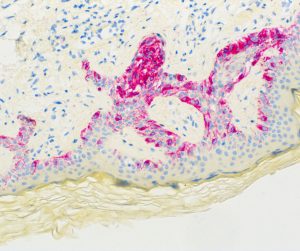
[5] Clarkson KS, Sturdgess IC and Molyneux AJ (2001): The usefulness of tyrosinase in the immunohistochemical assessment of melanocytic lesions: a comparison of the novel T311 antibody (anti-tyrosinase) with S-100, HMB45, and A103 (anti-melan-A). J Clin Pathol. 54(3):196-200.
[6] Kawakami Y, Eliyahu S, Delgado CH et al. (1994): Cloning of the gene coding for a shared human melanoma antigen recognized by autologous T cells infiltrating into tumor. Proc Natl Acad Sci U S A. 91(9):3515-9.
[7] Coulie PG, Brichard V, Van Pel A et al. (1994): A new gene coding for a differentiation antigen recognized by autologous cytolytic T lymphocytes on HLA-A2 melanomas. J Exp Med. 180(1):35-42.
[8] Yaziji H and Gown AM (2003): Immunohistochemical markers of melanocytic tumors. Int J Surg Pathol. 11(1):11-5.
[9] Ramos-Vara JA, Beissenherz ME, Miller MA et al. (2001): Immunoreactivity of A103, an antibody to Melan A, in canine steroid-producing tissues and their tumors. J Vet Diagn Invest. 13(4):328-32.
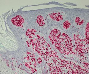
[10] Sheffield MV, Yee H, Dorvault CC et al. (2002): Comparison of five antibodies as markers in the diagnosis of melanoma in cytologic preparations. Am J Clin Pathol. 118(6):930-6.
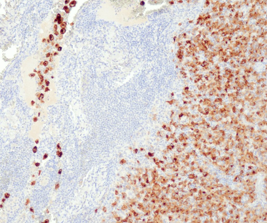
[11] Shidham VB, Qi D, Rao RN et al. (2003): Improved immunohistochemical evaluation of micrometastases in sentinel lymph nodes of cutaneous melanoma with ‚MCW melanoma cocktail‘–a mixture of monoclonal antibodies to MART-1, Melan-A, and tyrosinase. BMC Cancer. 3:15.